Payor Strategies
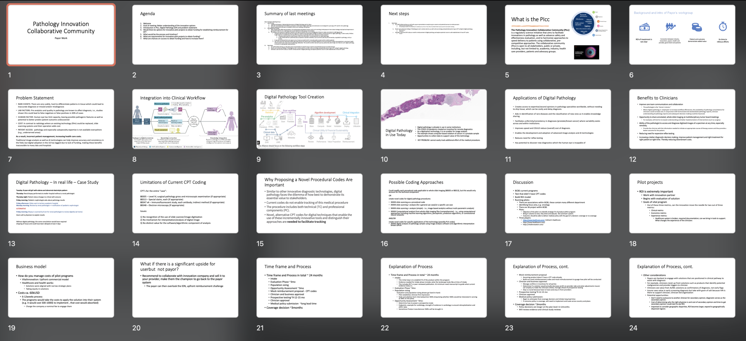
Financial sustainability is one of the key –if not the key– challenge of digital pathology and ML/AI applications in diagnostic pathology. Payors need conclusive and definitive clinical outcomes and clinical utility data. The return on investment for digital pathology solutions has not been conclusively shown. Other key challenges include that: clinical utility studies are costly but necessary and that effective integration of digital pathology into existing clinical decision-making guidelines is a long-term process. The workgroup is focusing on best practices to understand coverage determination pathways and is currently planning the following steps:
Identify evidence required for reimbursement
Identify endpoints needed to create evidence
Design a study to start building the evidence and request input from payors
Author a paper to incorporate digital pathology into practice guidelines, likely including above topics 1 and 2.
Key Elements, Next Steps, Timeline
The road to generating revenue for digital pathology should consider reimbursement for digital pathology devices as a whole, such as part of technology component of physician services fees or focus on individual diseases/indications.
Could consider bundled payments approach (e.g., DRG).
Need to include payors to understand pain points and educate medical directors of health plans.
Review non-pathology approaches to evidence generation.
Short term goal: identify optimal use case and evidence frameworks used by payors to inform development.
Midterm goal: develop evidence on shorter term endpoints while aligning with longer term follow up to inform outcomes.
Concerns & Problems
There is a high implementation cost and cost of storage and maintenance associated with use of digital pathology that is prohibitive to uptake. Labs need reimbursement to support use of digital pathology.
Efficiency gain from digital pathology may be interpreted as cost-savings by payors resulting in reduced rates of reimbursement. For example: Immunohistochemistry for certain use-cases might be able to be replaced (e.g., H. Pylori) resulting in decreased billable units?
Most payors have medical policy committees to conduct HTAs and evidence evaluation frameworks that are used to determine eligibility for coverage but these frameworks are not publicly available.
Ultimately evidence that digital pathology decreases cost and/or improves outcomes is needed, but who is going to pay for clinical utility studies?
Value Proposition
Workforce gap: Use of digital pathology will improve pathology resources during a period of decline in pathologists which overlaps with an anticipated growth in t in the field (40%) and increased need for pathology,
Economic impact:
Potential to decrease overall spending in healthcare through increased efficiency.
Potential to add value by less referrals, less confirmative testing, increased quality
Potential to decrease time to diagnosis
Clinical outcomes: potential to improve patient outcomes
Potential to provide more accurate diagnosis and thus improvement in response to therapy or delay in disease progression. Value of data: consider the importance of data to laboratories, payors, and patients as opposed to reimbursement as road to revenue
In summary:
Practice impact -> e.g. more efficient intervention -> improved patient experience
Clinical impact -> e.g. better treatment fit, higher response rate, fewer side effects -> improved patient outcome
Economic impact -> e.g. faster diagnosis -> reduction of downstream costs
Implications & Efforts
Efforts to advance evidence generation to support reimbursement will inform a road to revenue and monetization of ML/AI and more independent laboratories (agile players).
The field should be collecting real-world evidence from laboratories utilizing digital pathology to understand the extent of use and overall changes to use and outcomes during/after COVID-19 public health emergency.
Define key endpoints and outcomes: outline the key outcomes relevant to demonstration of the value-add of the technology.
Evaluate improvement in response to therapy or delay in disease progression are useful.
Consider the conduct benefit-risk assessments and development of cost-effectiveness data that can impact to relevant stakeholders including the hospital system and commercial payers.
Identify limitations of the current care paradigm: create a foundational understanding of the limitations of the current care paradigm so that the unique benefits of the technology can be highlighted.
A retrospective review of existing care decision-making and treatment patterns via chart review or claims analysis.
Pursue novel specific CPT codes, leveraging the new PLA coding system which allows for creation of codes specific to particular offerings/applications.
Build support for value-based payments: the evidence needed to support reimbursement in a value-based system will be different, requiring a move from viewing digital pathology as a reduction in cost to a source of value.
Define Clinical Utility of Digital Pathology Intervention: this can be achieved through an observational study which compares the selected endpoints from a program using the digital pathology technology to one without.
Relevant Resources
While the payor workgroup focuses on digital pathology (DP) and AI, the similarities of payor challenges in DP/AI mirrors that from comprehensive genotyping platforms (e.g. NGS sequencing and cfDNA). In this new white paper from Bruce Quinn, he provides an update on recent publications on plasma-based comprehensive genotyping, including health economics. He also reviews the status of PLA coding and Category I coding in detail. He argues that it is time for bringing the coding system up-to-date.
2021 Recap
-session at the “Pi” Annual Members Meeting that led to establishing a strategic agenda for the working group
-engagement with representatives from the AMA coding group to educate on the field of digital pathology and elicit feedback
-advisory sessions with a former Business Development Lead for one of BCBS Innovations program on a successful pilot program to develop evidence for coverage of innovative products
Current Projects
News & Updates
Relevant Publications
Current State of the Regulatory Trajectory for Whole Slide Imaging Devices in the USA
Date: May 2017
Authors: Abels et al.
Link: Pubmed
Implementation of whole slide imaging in surgical pathology: A value added approach
Date: 2011
Authors: Isaacs et al.
Link: Pubmed
Whole Slide Imaging Versus Microscopy for Primary Diagnosis in Surgical Pathology: A Multicenter Blinded Randomized Noninferiority Study of 1992 Cases (Pivotal Study)
Date: January 2018
Authors: Mukhopadhyay et al.
Link: Pubmed
Challenges in the Development, Deployment, and Regulation of Artificial Intelligence in Anatomic Pathology
Date: November 2020
Authors: Cheng et al.
Link: Pubmed
Clinical Application of Image Analysis in Pathology
Date: July 2020
Authors: Toby C Cornish
Link: Pubmed
Emerging role of deep learning-based artificial intelligence in tumor pathology
Date: April 2020
Authors: Jiang et al.
Link: Pubmed
Whole slide imaging: The futurescape of histopathology
Date: January-March 2021
Authors: Jayaram N Iyengar
Link: Pubmed
Whole slide imaging equivalency and efficiency study: experience at a large academic center
Date: July 2019
Authors: Hanna et al.
Link: Pubmed
Group Leaders
Esther Abels, MSc
Esther Abels has a background in bridging R&D, proof of concept, socio economics and pivotal clinical validation studies used for registration purposes in different geographies, for both pharma and biotech products. She brings to Visiopharm a wealth of regulatory and clinical experience specializing in bringing products to clinical utility. She played a crucial role in getting WSI devices reclassified in USA. Esther currently also leads the Digital Pathology Association (DPA) Regulatory and Standards Taskforce and FDA collaborations to drive regulatory and standard clarifications for interoperability and computational pathology in the field of digital pathology. She is also a co-founder of the Alliance for Digital Pathology. Esther holds a MSc in Biomedical Health Science from Radboud University Nijmegen.
Laura Lasiter, PhD
Laura Lasiter, PhD is Director of Health Policy at Friends of Cancer Research (Friends). Friends is an advocacy organization based in Washington, DC that drives collaboration among partners from every healthcare sector to power advances in science, policy, and regulation that speed life-saving treatments to patients. At Friends, Laura leads the strategic development and execution of Friends policy and legislative portfolio to affect evidence-based federal policy focused on modernized approaches to evidence generation in support of biomedical innovation.
Prior to joining Friends, Laura was a Congressional Science Fellow for the American Society of Microbiology/American Association for the Advancement of Science during which she covered the health portfolio of Senator Al Franken on the Senate HELP committee, primarily focused on issues relating to the FDA and prescription drug prices. She received her PhD in Biomedical Sciences from the University of Tennessee Health Science Center. Her doctoral work involved the characterization of the zoonotic potential of novel influenza viruses at St. Jude Children’s Research Hospital.
Jochen Lennerz, MD, PhD
Dr. Lennerz is board certified by the American Board of Pathology and the American Board of Medical Genetics. He joined the Massachusetts General Hospital Department of Pathology and Center for Integrated Diagnostics as a staff pathologist in 2014, and is an assistant professor at Harvard Medical School. Dr. Lennerz trained as a pathologist assistant in Berlin, Germany in 1994 and studied both medicine and molecular medicine at the University of Erlangen, Germany where he also received his MD and PhD. He completed his residency training in anatomic pathology in 2008, and a fellowship in molecular genetic pathology in 2009 at Washington University in St. Louis, MO. After completing a two-year gastrointestinal and liver pathology fellowship at Massachusetts General Hospital in 2011, he led a research group on biomarkers in lymphoma at Ulm University, Germany. His interests are tissue-based biomarkers, and financial sustainability of molecular genetic diagnostics.


